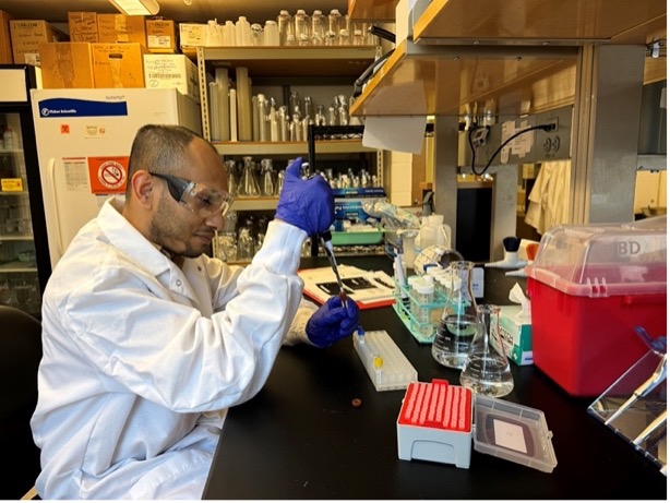
The Vision For Tomorrow Foundation is excited to share more details on the VFT funded research published in the medical journal “Science Translational Medicine”. The study, “Gene dosage manipulation alleviates manifestations of hereditary PAX6 haploinsufficiency in mice”, was published primarily by the University of Illinois Chicago (UIC) Department of Ophthalmology and Visual Sciences. Ali Djalilian, MD, co-author of the study, shared insights about the study findings and its potential impact on the Aniridia community with VFT.
“For nearly all of the genes in our DNA we have two copies, one which we inherit from our mother and one from our father. There are some genetic diseases where only one copy is normal and the other one is non-functional due to a mistake in the DNA,” said Djalilian. “The idea behind this study was to see if the normal copy can be enhanced to make up for the non-functional copy.”
The vast majority of Aniridia cases are caused by haploinsufficiency of the PAX6 gene. This means that one copy of the PAX6 gene is normal, but the other copy is either missing or mutated, making that copy non-functional. Since only one copy of the PAX6 gene is functional, only approximately 50 percent of the proteins necessary for normal function are produced. This reduced PAX6 expression in the eye contributes to the ocular problems faced by many with Aniridia, particularly issues with the cornea.
The main goal of the study was to determine if it is possible to stimulate the functional copy of PAX6 using an existing medicine to increase PAX6 expression in the eye. A secondary goal was to determine whether increasing PAX6 expression in the eye in this way would help to normalize eye development and function.
“Using a mouse model with aniridia, the team screened drugs that can enhance PAX6 and found a particular class of drugs known as MEK inhibitors that can stimulate PAX6 expression in the eye,” said Djaililan. “We tested this drug in newborn PAX6 deficient mice and found that either topical or oral administration enhanced PAX6 and partially normalized their eye development. In particular, they showed that the mice treated with topical MEK inhibitor had clearer corneas (less scarring) and could see better.”
Throughout the study, measurements were taken of the corneal epithelium, corneal stroma, corneal opacity, and size of the eyes. The researchers collaborated with scientists at the University of Virginia to test the retinal function and baseline vision of the mice.
The results in mice were very encouraging, particularly with the topical delivery. The most important results include:
- Wild-type mouse eyes showed no negatives from the treatment.
- Corneal epithelial thickness increased sizably. The epithelium is the outermost layer of the cornea, and aniridic epithelium tends to be thinner than normal. While this treatment did not bring the thickness back to normal, it did improve it significantly. The improvement in thickness persisted even 30 days after treatment ended.
- Corneal stromal thickness decreased significantly. The stroma is the middle and thickest layer of the cornea, and the aniridic stroma tends to be thicker than normal. Again, this treatment did not bring the thickness back to normal, but it did improve it significantly. This improvement persisted better in the topical group than in the oral group 30 days after treatment.
- Corneal opacity improved.
- Corneal epithelial PAX6 expression increased from 32 to 92 with topical application. Wild-type levels are 100, showing a notable improvement.
- Corneal epithelial differentiation was improved from 0 to 68. Again, wild-type levels are 100, so while not normal levels, this is a significant improvement. These results were still present 60 days after treatment ended in the topical application group.
- Corneal epithelial proliferation reduced from about 30 to around 1.5, which is close to the wild-type levels of 0.4.
- The mutant mouse model used for these experiments have very small eyes. After treatment, the eyes were significantly larger. Photos of the difference are striking and can be seen in this press release from UIC.
- Retina function was improved, even 60 days after treatment ended.
- Baseline vision improved significantly, even 90 days after treatment ended.
The study has important implications of the research for the Aniridia community.
- The results show repurposing drugs can work. Using existing medications to increase PAX6 expression in the eye is a legitimate avenue of research with great potential to help both the aniridic community and other genetic conditions resulting from haploinsufficiency.
- This approach could help prevent corneal keratopathy. Since keratopathy is a challenging problem to address clinically, finding preventative measures would be a significant accomplishment.
- This research is important because stimulating the functional copy of the PAX6 gene would work regardless of the type of mutation or deletion that caused aniridia.
- Past research has shown that, in addition to the eye, PAX6 expression also is required for development and maintenance of the brain, nervous system, olfactory system, and the pancreas. Theoretically, then, it is possible that increasing PAX6 expression through drug repurposing could also improve the function of those systems.
The study is a great example of researchers collaborating for maximum impact. James Lauderdale, PhD, University of Georgia, provided the small eye mouse model used in this study and researchers at the University of Virginia completed the testing of baseline vision and retinal function for the mice in this study. This type of collaboration was in practice at the 2019 John F. Anderson Symposium: PAX6 and Beyond. VFT was a sponsor of the forum which allowed for the scientific community to present aniridia research findings and facilitate collaboration on future studies.
“More studies and further understanding are needed before this approach can be tested in human patients, nonetheless, the study opens the possibility of using approved medications in some genetic diseases to enhance the function of a normal copy of a gene to compensate for the non-functional copy,” Djailian said. “This study was done in mice during their first month of life when the eye is still developing, but we need to still determine how this or similar drugs impacts the progression of the corneal disease if treatment is started beyond the developmental period.”
VFT is thankful for Dr. Djalilian, his team, and all the accomplished researchers that continue to pursue this promising research to help understand the challenges presented by aniridia. Please consider a donation to VFT to fuel more important scientific findings that can help to better understand and more effectively treat ocular complications related to aniridia and albinism.


